Haller Cell : Special ethmoidal air cells - How to Remember: MD/MS ... - The cells are variable in both size and number in the lateral mass of each of the ethmoid.
Haller Cell : Special ethmoidal air cells - How to Remember: MD/MS ... - The cells are variable in both size and number in the lateral mass of each of the ethmoid.. Mild mucosal thickening is seen in both ethmoid air cells and minimal in the frontal. Ethmoidal air cells that extend along the medial floor of the orbit. 'haller cells' — named after swedish anatomist albrecht von haller are abnormally migrated anterior or posterior ethmoid air cells that may pneumatize the roof of the maxillary sinus. Correspondence was rare in the same case between the. It is related to the orbital floor.
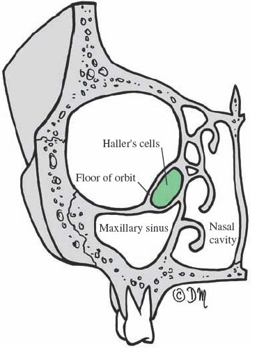
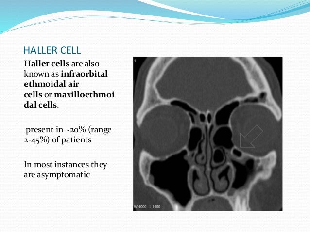
Ethmoid air cells, cells of ethmoid bone, cellulae ethmoideae osseae, bony ethmoidal cells, sinus haller, albrecht von. What does haller cell mean? The ethmoid sinuses or ethmoid air cells of the ethmoid bone are one of the four paired paranasal sinuses. Haller cells are infraorbital ethmoidal air cells that project from the maxillary sinus roof and the most inferior portion of the lamina papyracea. If haller cells were found, their capacity was determined.
Surgical Anatomy of the Paranasal Sinus | Ento Key from entokey.com Variation of posterior ethmoid cells located above the sphenoid sinus as a result of hyperpneumatization. Submitted 8 months ago by 158170863. This is formed by lateral and posterior pneumatization of the most posterior ethmoid cells over the sphenoid sinus. Concha bullosa and haller cell resection, maxillary sinus outflow obstruction, identification and removal of haller cells Meaning of haller cell medical term. Haller cells are located below the bulla ethmoidalis and extend beneath the floor of the orbit (figs 4 and 5). 'haller cells' — named after swedish anatomist albrecht von haller are abnormally migrated anterior or posterior ethmoid air cells that may pneumatize the roof of the maxillary sinus. They have been often implicated in several sinonasal diseases.
Haller cell assessment in preoperative planning becomes essential as pressure on the infraorbital nerve can trigger trigeminal nerve stimulation, especially in migraine with rhinogenic origin.
Haller cells are infraorbital ethmoidal air cells that project from the maxillary sinus roof and the most inferior portion of the lamina papyracea. Ethmoidal air cells that extend along the medial floor of the orbit. They can obstruct the outflow tract of the maxillary sinus and must be removed when there is pathology within. Cell — the basic structural and functional unit in people and all living things. Haller's cells are air cells located below the ethmoid bulla along the roof of the maxillary sinus. In the ct image haller cells are seen. Correspondence was rare in the same case between the. Squamous cell carcinoma of thyroid diagnostic & management dilemma. The cells are variable in both size and number in the lateral mass of each of the ethmoid. Variation of posterior ethmoid cells located above the sphenoid sinus as a result of hyperpneumatization. If orthopantomograms (opg) were taken the total number of haller cells was 64. Identification and management by sinusvideos on vimeo, the home for high quality videos and the people who love them. There is a close relation with the optic nerve.
Submitted 8 months ago by 158170863. Each cell in the human body there are 100 trillion cells in each of us contains the entire… … Variation of posterior ethmoid cells located above the sphenoid sinus as a result of hyperpneumatization. Correspondence was rare in the same case between the. Ethmoid air cells, cells of ethmoid bone, cellulae ethmoideae osseae, bony ethmoidal cells, sinus haller, albrecht von.
Endoscopic anatomy of nose ,paranasal sinus and anterior ... from image.slidesharecdn.com They can obstruct the outflow tract of the maxillary sinus and must be removed when there is pathology within. In the ct image haller cells are seen. What does haller cell mean? Intercolic anastomic artery, central anastomotic mesenteric artery, arcade of. At first an access to the. Meaning of haller cell medical term. It is related to the orbital floor. Mild mucosal thickening is seen in both ethmoid air cells and minimal in the frontal.
Haller cells are anterior ethmoid air cells located in the medial orbital floor immediately lateral to the haller cells are frequently seen as incidental findings in ct examinations of paranasal sinuses.
Ethmoid air cells, cells of ethmoid bone, cellulae ethmoideae osseae, bony ethmoidal cells, sinus haller, albrecht von. Named after albrecht von haller, a swiss anatomist. Meaning of haller cell medical term. There is an intraoperative relationship of the left haller cell to the maxillary sinus. What does haller cell mean? Haller cells lay posterosuperior to the natural maxillary os. It is related to the orbital floor. If orthopantomograms (opg) were taken the total number of haller cells was 64. Does anyone know anything about haller cells? Haller cell assessment in preoperative planning becomes essential as pressure on the infraorbital nerve can trigger trigeminal nerve stimulation, especially in migraine with rhinogenic origin. In the ct image haller cells are seen. Haller cells are infraorbital ethmoidal air cells that project from the maxillary sinus roof and the most inferior portion of the lamina papyracea. Haller cells are located below the bulla ethmoidalis and extend beneath the floor of the orbit (figs 4 and 5).
Squamous cell carcinoma of thyroid diagnostic & management dilemma. Haller cell assessment in preoperative planning becomes essential as pressure on the infraorbital nerve can trigger trigeminal nerve stimulation, especially in migraine with rhinogenic origin. The cells are variable in both size and number in the lateral mass of each of the ethmoid. Haller's cells, also known as infraorbital ethmoid cells are located at the medial roof of the maxillary sinus in the inferior most portion of the lamina papyracea. Submitted 8 months ago by 158170863.
Rt Haller cell fungal glue seen during fess surgery - YouTube from i.ytimg.com Intercolic anastomic artery, central anastomotic mesenteric artery, arcade of. Haller's cells are air cells located below the ethmoid bulla along the roof of the maxillary sinus. In the ct image haller cells are seen. Haller cells are infraorbital ethmoidal air cells that project from the maxillary sinus roof and the most inferior portion of the lamina papyracea. Ethmoidal air cells that extend along the medial floor of the orbit. They have been often implicated in several sinonasal diseases. Submitted 8 months ago by 158170863. Cell — the basic structural and functional unit in people and all living things.
Each cell in the human body there are 100 trillion cells in each of us contains the entire… …
They can obstruct the outflow tract of the maxillary sinus and must be removed when there is pathology within. This is formed by lateral and posterior pneumatization of the most posterior ethmoid cells over the sphenoid sinus. Bilateral haller air cells are seen. The cells are variable in both size and number in the lateral mass of each of the ethmoid. If haller cells were found, their capacity was determined. Haller's cells are air cells located below the ethmoid bulla along the roof of the maxillary sinus. Haller cell assessment in preoperative planning becomes essential as pressure on the infraorbital nerve can trigger trigeminal nerve stimulation, especially in migraine with rhinogenic origin. Ethmoid air cells, cells of ethmoid bone, cellulae ethmoideae osseae, bony ethmoidal cells, sinus haller, albrecht von. 'haller cells' — named after swedish anatomist albrecht von haller are abnormally migrated anterior or posterior ethmoid air cells that may pneumatize the roof of the maxillary sinus. Correspondence was rare in the same case between the. Haller's cells, also known as infraorbital ethmoid cells are located at the medial roof of the maxillary sinus in the inferior most portion of the lamina papyracea. Haller cells are anterior ethmoid air cells located in the medial orbital floor immediately lateral to the haller cells are frequently seen as incidental findings in ct examinations of paranasal sinuses. Each cell in the human body there are 100 trillion cells in each of us contains the entire… …
Haller cells are located below the bulla ethmoidalis and extend beneath the floor of the orbit (figs 4 and 5) haller. Haller cells are located below the bulla ethmoidalis and extend beneath the floor of the orbit (figs 4 and 5).